ORIGINAL ARTICLE

The use of lung ultrasound during the COVID-19 pandemic: A narrative review with specific focus on its role in pregnancy

Aly Youssef, MD, PhD1*, Marta Cavalera, MD1, Carlotta Azzarone, MD1, Carla Serra, MD, PhD2, Elena Brunelli, MD1, Paolo Casadio, MD, PhD1, Gianluigi Pilu, MD, PhD1

1Department of Obstetrics and Gynecology, Sant’Orsola Malpighi University Hospital, University of Bologna, Bologna, Italy

2Department of Organ Failure and Transplantation, Sant’Orsola Malpighi University Hospital, University of Bologna, Bologna, Italy

Abstract

The novel coronavirus disease (COVID-19) is a challenge to every health system. Unfortunately, it is unlikely that this pandemic will disappear soon. No health system, with its present resources and workflow, is capable enough to deal with a full-blown wave of this pandemic. Acquisition of specific new skills may be fundamental in delivering appropriate health care for our patients. The gold standard for diagnosis of the COVID-19 infection is real-time reverse transcription polymerase chain reaction. Radiological investigations (chest X-ray or high-resolution computerized tomography [CT]) can be helpful both for diagnosis and management, but they have many limitations. Ultrasound has been suggested as a reliable and accurate tool for assessing the lungs in COVID-19 patients. Lung ultrasound (LUS) can show specific signs of interstitial pneumonia, which is characteristic of COVID-19 pulmonary infection. In addition, nonradiologist specialists with experience in ultrasound can be trained on LUS with a relatively rapid learning curve. In pregnancy, LUS can be particularly useful due to the avoidance of exposure to ionizing radiation. In this review, we present the advantages, techniques, and limitations of the use of LUS during the COVID-19 pandemic, with specific focus on pregnancy.

Key words: lung ultrasound, COVID-19, SARS-CoV-2, pneumonia

*Corresponding author: Dr. Aly Youssef, MD, PhD, Department of Obstetrics and Gynecology, Sant’Orsola Malpighi University Hospital, University of Bologna, Bologna, Italy. Tel +39 051 214 4369. Email: [email protected]

Submitted: 13 July 2020; Accepted: 29 September 2020; Published: 15 October 2020.

DOI: 10.15586/jptcp.v27iSP1.743

©2020 Youssef A et al.

This article is distributed under the terms of the Creative Commons Attribution-Non Commercial 4.0 International License.This is an Open Access article distributed under the terms of the Creative Commons Attribution-NonCommercial-ShareAlike 4.0 International (CC BY-NC-SA 4.0). License (http://creativecommons.org/licenses/by-nc-sa/4.0/)

BACKGROUND

The novel coronavirus disease (COVID-19) is a global public health emergency. The disease is highly infectious and is characterized mainly by atypical pneumonia, caused by the SARS-CoV-2.1,2 Since the first case of COVID-19 pneumonia was reported in Wuhan, Hubei Province, China, in December 2019, the infection has spread rapidly to the rest of China and beyond. On 12 March 2020, on the basis of more than 20,000 confirmed cases and almost 1000 deaths in Europe, the World Health Organization announced the new coronavirus outbreak as a global pandemic.3 As to the day of writing of this review, more than 5 million confirmed cases and 330,000 deaths due to COVID-19 have been reported.4

No health system was appropriately prepared for the Tsunami-like surge of the COVID-19 pandemic, especially when the number of cases increased exponentially, as in the epicenters of the disease.5 In fact, even hospitals at the forefront have collapsed or ran the risk of collapse when overwhelmed by the exceptionally high number of COVID-19 cases.6 This pandemic had a profound impact on the departments that were responsible for the management of COVID-19 cases, and the consequences have reached almost every specialty in medicine. Almost all nonurgent visits and surgeries were halted in the vast majority of countries with COVID-19 cases.1,7,8 Furthermore, the consequences of the procedures and the steps taken to reduce the spread of infection and to redistribute the resources to deal with this pandemic have not always been appropriately assessed, and remain to be observed. Indeed, some estimates have raised serious concerns regarding the potential devastating consequences as a result of unavoidable shocks, health system collapse, or intentional choices made in response to the pandemic with regard to community morbidity and mortality.1,9

Acquisition of specific new skills may be fundamental in delivering appropriate health care for our patients. The gold standard for the diagnosis of COVID-19 infection is real-time reverse transcription polymerase chain reaction (RT-PCR). Radiological investigations (chest X-ray or high-resolution CT) can be helpful both for diagnosis and management, but they have many limitations. Ultrasound has been suggested as a reliable and accurate tool for assessing the lungs in COVID-19 patients.

COVID-19 AND PREGNANCY

The presence of COVID-19 in a pregnant patient raises concerns, as other types of coronaviruses were frequently associated with an increased risk of adverse outcomes in comparison with the general population of the same age.1012 Fortunately, to date, there is no evidence that pregnant women are more susceptible to COVID-19 infection, nor to complications and mortality as a result of the disease.1316 Moreover, there is no evidence of vertical transmission (maternal-to-neonatal) of COVID-19 infection when the maternal infection manifests in the third trimester and of severe adverse outcomes in neonates.17 Another comforting data are the absence of convicing evidence on intrapartum transmission of COVID-19 via vaginal delivery to the neonates.15,1719 The most common obstetrical complication to COVID-19 seems to be preterm delivery and the high risk of Cesarean delivery.15 Both do not seem to be due to the disease itself, but due to iatrogenic intervention, and more data are needed to understand the relationship between COVID-19 and the mode of delivery and perinatal outcomes.

DIAGNOSIS OF COVID-19

  1. The role of clinical assessment: Initially, a history of traveling to areas with a high number of cases, “hot spots,” was often used to define a patient’s risk. Now, in most areas, due to the wide spread of the disease, it is acknowledged that clinical assessment is the main tool to define suspected cases. The most common clinical findings in cases of COVID-19 infection include the following: fever, dry cough, myalgia, fatigue, and dyspnea.20 Less common clinical features are headache, dizziness, abdominal pain, diarrhea, nausea, and vomiting. Common laboratory findings are lymphopenia, prolonged prothrombin time, and elevated lactate dehydrogenase.21 However, a large proportion of cases positive for SARS-CoV-2 will have very mild or no symptoms.2224 A recent cochrane review concluded that the individual signs and symptoms of COVID-19 appear to have very poor diagnostic properties.25

  2. Reverse transcription polymerase chain reaction: The gold standard for COVID-19 infection diagnosis remains the detection of SARS-CoV-2 RNA in nasopharynx samples by RT-PCR.26 Various concerns remain regarding the RT-PCR. These include shortage in kit supplies, especially in low-resource countries. Another major limitation is the rate of false negatives. This may be due to irregular sampling, laboratory error, insufficient viral material in the specimen, improper extraction of nucleic acid from clinical materials, and contamination and technical problems.27,28 The screening policies by RT-PCR and the number of swabs per 1000 citizens remain extremely heterogenous among countries and even regions in the same country.29 COVID-19 is highly contagious; therefore, early detection and diagnosis are of utmost importance to isolate suspected cases to control the outbreak. The above-mentioned challenges in the identification of pathogens by RT-PCR made many clinicians propose radiological investigations as a diagnostic method for identifying COVID-19 patients.30

  3. Radiological investigations

    1. Computerized tomography

      Many studies have demonstrated that CT can be a very useful tool for screening, diagnosis, and follow-up of COVID-19 cases.31 The most frequent CT abnormalities found in patients diagnosed with COVID-19 are ground glass opacity, consolidation, and interlobular septal thickening.32 Lung lesions are usually multiple, and the most involved lung segments are the lower lobes bilaterally. In spite of the several advantages offered by CT, it has some limitations. These include the cost, the high dose of radiation, the need to move the patient across various buildings to be examined by different health personnel, thereby increasing the risk of contamination. In fact, radiology societies have advised against the systematic use of CT and have suggested reducing the number of chest X-rays in COVID-19 patients, to minimize the exposure of health care providers to this viral infection.33 Considering the need to screen and monitor as many patients as possible during the pandemic, lung ultrasound (LUS) was performed with the aim of identifying quickly those patients more likely to be affected by COVID-19.

    2. Lung ultrasound

      • Advantages and limitations of LUS (Table 1)

      • Accuracy of LUS in the diagnosis of pneumonia

        Many studies have found that LUS is reliable in lung assessment. It has been demonstrated that LUS is sensitive, specific, and reproducible for diagnosing the main lung pathologic entities in patients with Acute Respiratory Distress Syndrome (ARDS). It has thus been advocated that LUS can be considered an attractive alternative to bedside chest X-ray and thoracic computed tomography (CT).44

        In addition, LUS seems to be quite reliable in the diagnosis of pneumonia. In a study on patients with community-acquired pneumonia, LUS was found to be helpful in diagnosis with better accuracy in comparison with chest X-ray, using chest CT scan as the gold standard.45 The sensitivity and specificity of LUS was 95 and 90%, respectively. Similar results were found by another multicenter prospective study on patients with various respiratory diseases.46 The authors concluded that LUS should be part of routine diagnostic algorithm of respiratory diseases.

      • Usefulness of LUS in the diagnosis and management of COVID-19

        Few data exist on the reliability of LUS in the diagnosis of COVID-19 pneumonia. The diagnostic efficacy seems to depend on the severity of lung involvement, with higher accuracy in more severe cases.47 As this kind of pneumonia is more likely to start from the lateral zone of the lung, close to the pleura, LUS can more effectively demonstrate the pathologic and dynamic changes of the lung condition.48 This characteristic makes LUS an important tool for monitoring the evolution of disease. LUS has high diagnostic accuracy and fewer infection control implications due to decreased patient transportation between various hospital departments. Furthermore, the LUS score provides a degree of objectivity to quantifying the level of involvement, as a comprehensive whole organ assessment.49,50 There is growing evidence suggesting that LUS is accurate both in terms of the diagnosis and the prediction of prognosis and the evolution of lung disease in COVID-19.35,39,40,42,5154 Indeed, it has been suggested that LUS may be used as a primary imaging modality for pulmonary assessment, thereby reducing the use of chest X-ray in COVID-19 patients.55 LUS can reliably differentiate COVID-19 pneumonia from community-acquired pneumonia.56

TABLE 1. Advantages and Limitations of LUS

Advantages Limitations
  • The ability to perform a point-of-care evaluation

  • Safety and the absence of ionizing radiation

  • Reduced costs

  • The ease of repeating the examination

  • The absence of a need to move the patient from the ward of admission, thereby limiting the risk of spread of the infection.34,35

  • An ultrasound-based approach in the management of COVID-19 has proven to help in decision-making, reduce the number of chest X-rays and thoracic CT scans, thereby reducing the number of health care providers exposed to possible contamination and sparing personal protective equipment.3638

  • In pregnancy, LUS can be particularly useful due to the avoidance of the exposure to ionizing radiation.

  • The ability to teach it relatively rapidly in comparison with other diagnostic and follow-up tools for COVID-19.34,39 In its full-blown wave, most countries have recruited physicians from a wide range of medical specialties to help in the management of the pandemic. Ultrasound is already used in many medical fields (obstetrics, gynecology, cardiology, pediatrics, etc…). This has led to a rising interest in the application of LUS by many specialists who do not routinely perform LUS in their daily practice, especially those who already have experience in ultrasound for other indications.4043

  • Ultrasound cannot usually detect lesions that are deep and intrapulmonary. In a normal lung, the only lung structure that can be visualized is the pleura. In case of any pathology involving the pleura and/or the lung tissue adjacent to the pleural line, LUS can be useful. In case of isolated deep lesions, such as lung tumors, ultrasound is unlikely to detect any abnormal findings.45,46

  • LUS is operator-dependent with more experienced sonographers having higher diagnostic accuracy.47

  • LUS is also patient-dependent. In fact, obese patients may be more difficult to examine due to the thickness of their soft tissues.46 The second limitation of LUS is its lower accuracy compared to CT scan.

LUS, lung ultrasound; CT, computerized tomography.

TECHNICAL ASPECTS OF LUS

  1. Before you start: safety procedure awareness

    When planning an ultrasound examination in the context of the COVID-19 pandemic, some crucial aspects should be considered in addition to the routine procedures. These include mainly:

    • the means to protect the patient and the ultrasound providers.

    • the methods to prepare and clean the ultrasound room and equipment. A recent detailed position statement covering both points has been published by the International Society of Ultrasound in Obstetrics and Gynecology.57

  2. The transducer

    LUS examination can be performed with almost all types of ultrasound machines. Different types of probes can be used: convex, linear, or phased-array.58

  3. Machine settings

    It is crucial to standardize the machine settings in order to optimize diagnosis and avoid the wrong interpretation of findings in the case of follow-up of the same patients.59

    Ultrasound gain should be reduced in order to increase the contrast and highlight the hyperechoic signs. It is recommended to remove contrast enhancement techniques.

  4. Patient position

    LUS can be performed in supine, sitting, and lateral positions.

  5. Lung areas to be examined

    Both hemithoraxes should be covered, anteriorly, laterally, and posteriorly, from basal to upper zones. Many approaches have been suggested.60,61

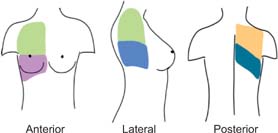
    One of the most practical and used approaches can be seen in Figure 1. Each hemithorax is divided into six regions with the aid of anatomical landmarks set by anterior and posterior axillary lines: two anterior, two lateral, and two posterior regions (Figure 2). Each region should be examined in sagittal and axial views.

FIG 1. Areas to be examined using lung ultrasound. Each hemithorax is subdivided into six areas by the anterior and posteior axillary lines.

FIG 2. The six regions to be examined by lung ultraound.

NORMAL AND ABNORMAL FINDINGS

  1. Normal findings

    Pleural line (Figure 3): in a normal lung, only the pleural line can be seen. This appears as a hyperechoic and sliding line, moving forward and backward with ventilation. This is the result of the movement of the visceral pleura against the parietal pleura during the respiratory cycle.

    A-lines (Figure 4): these appear as motionless and regularly spaced horizontal lines under the pleural line. These correspond to normal reverberation artefact of the pleural line. A-lines and lung sliding define normal aeration.62

  2. Abnormal findings

    Abnormal pleural line (Figure 5): in inflammatory lung disease, such as early COVID-19 pneumonia, the pleura line is usually irregular and blurred.39

    Well-separated B-lines (Figure 6): one of the hallmarks of acute lung injury is increased extravascular lung water (EVLW), which extends to the lung peripheries. This produces vertical ultrasound artefacts resulting from abnormal gas–tissue interface. B-lines start from the pleural line and “shine” vertically to erase A-lines and reach the end of the screen. The presence of B-lines reflects moderate lung aeration loss resulting from interstitial syndrome. B-lines are often sparse and separated.

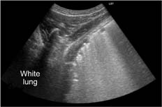
    Coalescent B-lines (white lung; Figure 7): B-lines may amalgamate, giving the image of “coalescent” B-lines arising from the pleura and spreading to the edge of the screen, denoting more severe lung edema.

FIG 3. Normal pleural line.
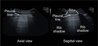
FIG 4. A-lines.

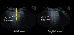
FIG 5. Thickened and irregular pleural line.
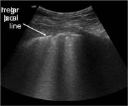
FIG 6. B-lines.

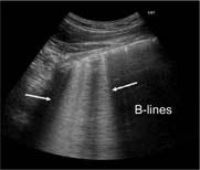
FIG 7. Coalescent B-lines (white lung).

NOTE: “The presence of B-lines denotes interstitial lung disease but does not indicate the pertinent etiology. However, we think that in the context of a COVID-19 pandemic, patients with LUS findings suggestive of interstitial lung disease should be considered to have COVID-19 pneumonia until proven otherwise.”

Lung consolidation (Figure 8): this is due to massive aeration loss. Lungs acquire a tissue-like echotexture (hypoechoic appearance). Lung consolidation usually has a superficial boundary at the level of the pleural line and a deep boundary, which is usually irregular with the aerated lung or regular in the case of whole lobe involvement.

Air bronchogram (Figure 9): sometimes, hyperechoic punctiform images may be seen within the consolidation corresponding to the air-filled bronchi. This is also called air bronchograms. During inspiration, these hyperechoic punctiform images may be reinforced due to penetration of gas into the bronchial tree of the consolidation (the so-called dynamic bronchogram).

FIG 8. Lung consolidation.

FIG 9. Lung consolidation with air bronchogram.

THE LUNG ULTRASOUND SCORE

Qualitative assessment has been proposed by some authors, while others have suggested the use of a quantitative score.52,62 By attributing each examined region a score per normal or abnormal findings, LUS is the sum of the scores of all examined areas.

Scoring Procedures52: Each examined area is given a score from 0 to 3. Score 0: The pleural line is continuous and regular. Horizontal normal artifacts are present. Score 1: The pleural line is indented, with vertical areas of white lung visible below it. Score 2: The pleural line is broken, with some consolidated areas visible with associated areas of white lung under the breaking point. Score 3: The scanned area shows dense and a largely extended white lung which can also be associated with larger consolidations. At the end of the procedure, the operator will note the highest score obtained for each examined area.

A quantitative score can improve reproducibility. Furthermore, it can be extremely useful in the documentation of the progression or the improvement of lung involvement. Due to its simplicity and the various above-mentioned advantages, some authors tried to examine the accuracy of LUS in screening for COVID-19. It is important to note that while LUS seems accurate and reliable in diagnosing patients with COVID-19 pneumonia, the available data demonstrate that it is unlikely to be a useful screening tool in asymptomatic patients.6365

LUS IN PREGNANCY

In addition to the already mentioned advantages, LUS has further benefits in pregnant women. Avoiding ionizing radiation is particularly beneficial in pregnancy to prevent fetal exposure. Furthermore, almost all women perform several fetal ultrasound examinations in pregnancy.66

This means that a potential LUS would be practically at zero cost. Various studies on pregnant women showed that LUS is highly accurate in the diagnosis of COVID-19 pneumonia. LUS seems accurate also for the longitudinal monitoring of the pathology in pregnant women and provides a safe and accurate alternative to CT and chest X-ray.43,54,67

To sum up, LUS is a safe, inexpensive, and reliable tool for the assessment of COVID-19. Training various specialists with some experience of ultrasound in other fields seems feasible and efficient, especially in the peak or in preparation for the peak of the COVID-19 outbreak. In spite of its several advantages, it is unlikely that LUS will replace CT in a clinical setting; however, it will probably provide a useful tool in the armamentarium of the clinicians managing COVID-19 patients.

CONCLUSIONS

LUS is a reliable tool for assessing the lungs in COVID-19 patients. LUS can show specific signs of interstitial pneumonia, which is characteristic of COVID-19 pulmonary infection. LUS has many advantages including its relatively rapid learning curve, the bedside applicability, and the absence of ionizing radiation. In pregnancy, LUS can be particularly useful due to the avoidance of the exposure of fetuses to ionizing radiation.

CONFLICT OF INTEREST

The authors report no conflict of interest.

REFERENCES

1. Casadio P, Youssef A, Arena A, et al. Increased rate of ruptured ectopic pregnancy in COVID-19 pandemic: Analysis from the North of Italy. Ultrasound Obstet Gynecol 2020;56(2):289. 10.1002/uog.22126

2. Arons MM, Hatfield KM, Reddy SC, et al. Presymptomatic SARS-CoV-2 infections and transmission in a skilled nursing facility. N Engl J Med 2020;382(22):2081–90. 10.1056/NEJMoa2008457

3. World Health Organization. Coronavirus disease (COVID-19) pandemic. https://www.who.int/emergencies/diseases/novel-coronavirus-2019 (accessed on May 20, 2020).

4. Coronavirus COVID-19 global cases. Johns Hopkins CSSE. https://gisanddata.maps.arcgis.com/apps/opsdashboard/index.html#/bda7594740fd40299423467b48e9ecf6 (accessed on May 21, 2020).

5. Maniscalco P, Poggiali E, Quattrini F, et al. The deep impact of novel CoVID-19 infection in an Orthopedics and Traumatology Department: The experience of the Piacenza Hospital. Acta Biomed 2020;91(2):97–105.

6. Senni M. COVID-19 experience in Bergamo, Italy. Eur Heart J 2020;41(19):1783–4. 10.1093/eurheartj/ehaa279

7. Diaz A, Sarac BA, Schoenbrunner AR, et al. Elective surgery in the time of COVID-19. Am J Surg 2020;219(6):900–2. 10.1016/j.amjsurg.2020.04.014

8. Iacobucci G. Covid-19: All non-urgent elective surgery is suspended for at least three months in England. BMJ 2020;368:m1106. 10.1136/bmj.m1106

9. Roberton T, Carter ED, Chou VB, et al. Early estimates of the indirect effects of the COVID-19 pandemic on maternal and child mortality in low-income and middle-income countries: A modelling study. Lancet Glob Health 2020;8(7):e901–8. 10.1016/S2214-109X(20)30229-1

10. Di Mascio D, Khalil A, Saccone G, et al. Outcome of Coronavirus spectrum infections (SARS, MERS, COVID 1-19) during pregnancy: A systematic review and meta-analysis. Am J Obstet Gynecol MFM 2020. May; 2(2): 100107. 10.1016/j.ajogmf.2020.100107

11. Di Mascio D, Sen C, Saccone G, et al. Risk factors associated with adverse fetal outcomes in pregnancies affected by Coronavirus disease 2019 (COVID-19): A secondary analysis of the WAPM study on COVID-19. J Perinat Med 2020. (epub ahead of print)

12. Di Mascio D, COVID Wwgo. Maternal and perinatal outcomes of pregnant women with SARS-COV-2 infection. Ultrasound Obstet Gynecol 2020.(epub ahead of print)

13. Poon LC, Yang H, Dumont S, et al. ISUOG Interim Guidance on coronavirus disease 2019 (COVID-19) during pregnancy and puerperium: Information for healthcare professionals—An update. Ultrasound Obstet Gynecol 2020;55(6):848–62. 10.1002/uog.22061

14. Poon LC, Yang H, Lee JCS, et al. ISUOG Interim Guidance on 2019 novel coronavirus infection during pregnancy and puerperium: Information for healthcare professionals. Ultrasound Obstet Gynecol 2020;55(5):700–8. 10.1002/uog.22013

15. Della Gatta AN, Rizzo R, Pilu G, Simonazzi G. Coronavirus disease 2019 during pregnancy: A systematic review of reported cases. Am J Obstet Gynecol 2020;223(1):36–41. 10.1016/j.ajog.2020.04.013

16. ISUOG Safety Committee Position Statement on safe performance of obstetric and gynecological scans and equipment cleaning in context of COVID-19. Ultrasound Obstet Gynecol 2020;55(5):709–12. 10.1002/uog.22027

17. Chen H, Guo J, Wang C, et al. Clinical characteristics and intrauterine vertical transmission potential of COVID-19 infection in nine pregnant women: A retrospective review of medical records. Lancet 2020;395(10226):809–15. 10.1016/S0140-6736(20)30360-3

18. Yang H, Wang C, Poon LC. Novel coronavirus infection and pregnancy. Ultrasound Obstet Gynecol 2020;55(4):435–7. 10.1002/uog.22006

19. Khan S, Peng L, Siddique R, et al. Impact of COVID-19 infection on pregnancy outcomes and the risk of maternal-to-neonatal intrapartum transmission of COVID-19 during natural birth. Infect Control Hosp Epidemiol 2020;41(6):748–50. 10.1017/ice.2020.84

20. Fu L, Wang B, Yuan T, et al. Clinical characteristics of coronavirus disease 2019 (COVID-19) in China: A systematic review and meta-analysis. J Infect 2020;80(6):656–65. 10.1016/j.jinf.2020.03.041

21. Wang D, Hu B, Hu C, et al. Clinical characteristics of 138 hospitalized patients with 2019 novel Coronavirus-infected pneumonia in Wuhan, China. JAMA 2020. Mar 17;323(11):1061-1069. 10.1001/jama.2020.1585

22. Vintzileos WS, Muscat J, Hoffmann E, et al. Screening all pregnant women admitted to labor and delivery for the virus responsible for coronavirus disease 2019. Am J Obstet Gynecol 2020. Aug;223(2):284-286. 10.1016/j.ajog.2020.04.024. Epub 2020 Apr 26.

23. Sutton D, Fuchs K, D’Alton M, Goffman D. Universal screening for SARS-CoV-2 in women admitted for delivery. N Engl J Med 2020;382(22):2163–4. 10.1056/NEJMc2009316

24. Treibel TA, Manisty C, Burton M, et al. COVID-19: PCR screening of asymptomatic health-care workers at London hospital. Lancet 2020;395(10237):1608–10. 10.1016/S0140-6736(20)31100-4

25. Struyf T, Deeks JJ, Dinnes J, et al. Signs and symptoms to determine if a patient presenting in primary care or hospital outpatient settings has COVID-19 disease. Cochrane Database Syst Rev 2020;7:CD013665. 10.1002/14651858.CD013665

26. Corman VM, Landt O, Kaiser M, et al. Detection of 2019 novel coronavirus (2019-nCoV) by real-time RT-PCR. Euro Surveill 2020;25(3). 10.2807/1560-7917.ES.2020.25.3.2000045

27. Li Y, Yao L, Li J, et al. Stability issues of RT-PCR testing of SARS-CoV-2 for hospitalized patients clinically diagnosed with COVID-19. J Med Virol 2020;92(7):903–8. 10.1002/jmv.25786

28. Ai T, Yang Z, Hou H, et al. Correlation of Chest CT and RT-PCR Testing in Coronavirus Disease 2019 (COVID-19) in China: A report of 1014 cases. Radiology 2020 Aug; 296(2):E32-E40. 10.1148/radiol.2020200642. Epub 2020 Feb 26.

29. www.worldometers.info/coronavirus/ (accessed on May 29, 2020).

30. Yang W, Sirajuddin A, Zhang X, et al. The role of imaging in 2019 novel coronavirus pneumonia (COVID-19). Eur Radiol 2020. Sep;30(9):4874-4882. 10.1007/s00330-020-06827-4. Epub 2020 Apr 15.

31. Wang K, Kang S, Tian R, et al. Imaging manifestations and diagnostic value of chest CT of coronavirus disease 2019 (COVID-19) in the Xiaogan area. Clin Radiol 2020;75(5):341–7. 10.1016/j.crad.2020.03.004

32. Wu J, Wu X, Zeng W, et al. Chest CT findings in patients with coronavirus disease 2019 and its relationship with clinical features. Invest Radiol 2020;55(5):257–61. 10.1097/RLI.0000000000000670

33. Simpson S, Kay FU, Abbara S, et al. Radiological society of North America expert consensus statement on reporting chest CT findings related to COVID-19. Endorsed by the Society of Thoracic Radiology, the American College of Radiology, and RSNA—Secondary Publication. J Thorac Imaging 2020;35(4):219–27. 10.1097/RTI.0000000000000524

34. Buonsenso D, Moro F, Inchingolo R, et al. Effectiveness of rapid lung ultrasound training program for gynecologists and obstetricians managing pregnant women with suspected COVID-19. Ultrasound Obstet Gynecol. 2020 Jul 56(1):110–111. 10.1002/uog.22066.

35. Buonsenso D, Pata D, Chiaretti A. COVID-19 outbreak: Less stethoscope, more ultrasound. Lancet Respir Med 2020;8(5):e27. 10.1016/S2213-2600(20)30120-X

36. Mongodi S, Orlando A, Arisi E, et al. Lung ultrasound in patients with acute respiratory failure reduces conventional imaging and health care provider exposure to COVID-19. Ultrasound Med Biol 2020;46(8):2090–3. 10.1016/j.ultrasmedbio.2020.04.033

37. Vetrugno L, Bove T, Orso D, et al. Our Italian experience using lung ultrasound for identification, grading and serial follow-up of severity of lung involvement for management of patients with COVID-19. Echocardiography 2020;37(4):625–7. 10.1111/echo.14664

38. Smargiassi A, Soldati G, Borghetti A, et al. Lung ultrasonography for early management of patients with respiratory symptoms during COVID-19 pandemic. J Ultrasound. 2020 Jul 7:1–8. 10.1007/s40477-020-00501-7. Online ahead of print.

39. Moro F, Buonsenso D, Moruzzi MC, et al. How to perform lung ultrasound in pregnant women with suspected COVID-19. Ultrasound Obstet Gynecol 2020;55(5):593–8. 10.1002/uog.22028

40. Kiamanesh O, Harper L, Wiskar K, et al. Lung ultrasound for cardiologists in the time of COVID-19. Can J Cardiol 2020. Jul;36(7):1144–47. 10.1016/j.cjca.2020.05.008. Epub 2020 May 19.

41. Lugnan C, Tommasini V, Furlanis G, et al. Lung ultrasonography in COVID-19: A game changer in the stroke unit? Eur J Neurol 2020. May 25;10.1111/ene.14352. Online ahead of print.

42. Musolino AM, Supino MC, Buonsenso D, et al. Lung ultrasound in children with COVID-19: Preliminary findings. Ultrasound Med Biol 2020;46(8):2094–8. 10.1016/j.ultrasmedbio.2020.04.026

43. Youssef A, Serra C, Pilu G. Lung ultrasound in the coronavirus disease 2019 pandemic: A practical guide for obstetricians and gynecologists. Am J Obstet Gynecol 2020;223(1):128–31. 10.1016/j.ajog.2020.05.014

44. Lichtenstein D, Goldstein I, Mourgeon E, et al. Comparative diagnostic performances of auscultation, chest radiography, and lung ultrasonography in acute respiratory distress syndrome. Anesthesiology 2004;100(1):9–15. 10.1097/00000542-200401000-00006

45. Ye X, Xiao H, Chen B, Zhang S. Accuracy of lung ultrasonography versus chest radiography for the diagnosis of adult community-acquired rneumonia: Review of the literature and meta-analysis. PLoS One 2015;10(6):e0130066. 10.1371/journal.pone.0130066

46. Rinaldi L, Milione S, Fascione MC, et al. Relevance of lung ultrasound in the diagnostic algorithm of respiratory diseases in a real-life setting: A multicentre prospective study. Respirology 2020;25(5):535–42. 10.1111/resp.13659

47. Lu W, Zhang S, Chen B, et al. A clinical study of noninvasive assessment of lung lesions in patients with coronavirus disease-19 (COVID-19) by bedside ultrasound. Ultraschall Med 2020;41(3):300–7. 10.1055/a-1154-8795

48. Zhang Z, Ren B, Fan H, et al. The role of lung ultrasound in the assessment of novel coronavirus pneumonia. J Cardiothorac Vasc Anesth 2020. Oct;34(10):2851–2854.

49. Smith MJ, Hayward SA, Innes SM, Miller ASC. Point-of-care lung ultrasound in patients with COVID-19—A narrative review. Anaesthesia 2020. Aug;75(8):1096–1104. 10.1111/anae.15082. Epub 2020 Apr 28

50. Bouhemad B, Brisson H, Le-Guen M, et al. Bedside ultrasound assessment of positive end-expiratory pressure-induced lung recruitment. Am J Respir Crit Care Med 2011;183(3):341–7. 10.1164/rccm.201003-0369OC

51. Joob B, Wiwanitkit V. Lung ultrasound in pregnant women with suspicion of COVID-19. Ultrasound Obstet Gynecol 2020;56(1):123–4. 10.1002/uog.22091

52. Soldati G, Smargiassi A, Inchingolo R, et al. Proposal for international standardization of the use of lung ultrasound for patients with COVID-19: A simple, quantitative, reproducible method. J Ultrasound Med 2020;39(7):1413–19. 10.1002/jum.15285

53. Yassa M, Birol P, Mutlu AM, et al. Lung ultrasound can influence the clinical treatment of pregnant women with COVID-19. J Ultrasound Med 2020. Jun 1;10.1002/jum.15367

54. Buonsenso D, Raffaelli F, Tamburrini E, et al. Clinical role of lung ultrasound for the diagnosis and monitoring of COVID-19 pneumonia in pregnant women. Ultrasound Obstet Gynecol 2020. Jul;56(1):106–109. 10.1002/uog.22055

55. Moller-Sorensen H, Gjedsted J, Lind Jorgensen V, Lindskov Hansen K. COVID-19 assessment with bedside lung ultrasound in a population of intensive care patients treated with mechanical ventilation and ECMO. Diagnostics (Basel) 2020;10(7):447. 10.3390/diagnostics10070447.

56. Tan G, Lian X, Zhu Z, et al. Use of lung ultrasound to differentiate coronavirus disease 2019 (COVID-19) pneumonia from community-acquired pneumonia. Ultrasound Med Biol 2020. Oct;46(10):2651–2658. 10.1016/j.ultrasmedbio.2020.05.006

57. Poon LC, Abramowicz JS, Dall’Asta A, et al. ISUOG Safety Committee Position Statement on safe performance of obstetric and gynecological scans and equipment cleaning in context of COVID-19. Ultrasound Obstet Gynecol 2020;55(5):709–12. 10.1002/uog.22027

58. Gargani L, Volpicelli G. How I do it: Lung ultrasound. Cardiovasc Ultrasound 2014;12:25. 10.1186/1476-7120-12-25

59. Rouby JJ, Arbelot C, Gao Y, et al. Training for lung ultrasound score measurement in critically ill patients. Am J Respir Crit Care Med 2018;198(3):398–401. 10.1164/rccm.201802-0227LE

60. Lichtenstein DA, Mezière GA. Relevance of lung ultrasound in the diagnosis of acute respiratory failure: The BLUE protocol. Chest. 2008;134(1):117–25. 10.1378/chest.07-2800

61. Via G, Storti E, Gulati G, et al. Lung ultrasound in the ICU: From diagnostic instrument to respiratory monitoring tool. Minerva Anestesiol 2012;78(11):1282–96.

62. Bouhemad B, Mongodi S, Via G, Rouquette I. Ultrasound for “lung monitoring” of ventilated patients. Anesthesiology 2015;122(2):437–47. 10.1097/ALN.0000000000000558

63. Scheier E, Guri A, Balla U. Lung ultrasound cannot be used to screen for Covid-19 in children. Eur Rev Med Pharmacol Sci 2020;24(9):4623–4.

64. Youssef A, Brunelli E, Azzarone C, et al. Lung ultrasound is not a useful screening tool for severe acute respiratory syndrome coronavirus 2 in pregnant women: A pilot study. J Ultrasound Med 2020. 10.1002/jum.15451.

65. Kalafat E, Yassa M, Koc A, et al. Utility of lung ultrasound assessment for probable SARS-CoV-2 infection during pregnancy and universal screening of asymptomatic individuals. Ultrasound Obstet Gynecol 2020. Oct;56(4):624–626. 10.1002/uog.23099

66. Lowe SW, Pruitt RH, Smart PT, Dooley RL. Routine use of ultrasound during pregnancy. Nurse Pract 1998;23(10):60, 63–6, 71. 10.1097/00006205-199810000-00003

67. Inchingolo R, Smargiassi A, Moro F, et al. The diagnosis of pneumonia in a pregnant woman with coronavirus disease 2019 using maternal lung ultrasound. Am J Obstet Gynecol 2020;223(1):9–11. 10.1016/j.ajog.2020.04.020